

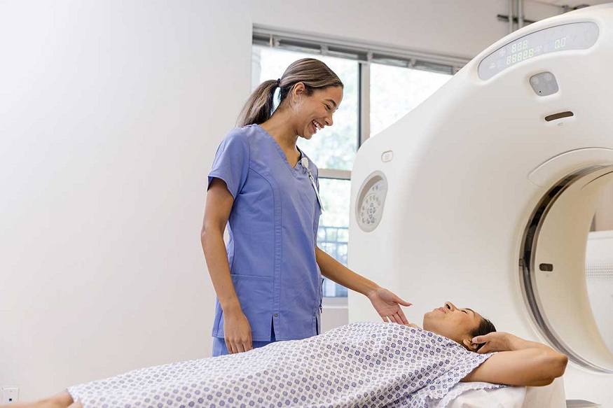
Magnetic Resonance Imaging (MRI) has become one of the most advanced and reliable diagnostic technologies available in modern healthcare. As Kasey McKillip points out, it enables physicians to obtain exceptionally detailed images of organs, muscles, joints, nerves, blood vessels, and other internal structures without exposing patients to ionizing radiation.
Kasey McKillip briefly discusses how MRI professionals prepare patients for a scan
While the technology is remarkably safe, the MRI environment is unlike any other medical imaging setting because it relies on an extremely powerful magnetic field that remains active at all times. For many individuals, the thought of entering an MRI scanner can feel overwhelming, especially if it is their first experience. This is why MRI technologists and radiology professionals follow a comprehensive preparation process before every examination. Their goal is to ensure patient safety, produce the highest quality diagnostic images, and make the overall experience as comfortable and stress-free as possible.
Read More: Maintaining a healthy lifestyle can lead to better medical test results.
The preparation process begins with an extensive safety evaluation, which is considered the most critical stage before an MRI examination. Since the scanner contains a powerful magnet that cannot simply be switched off, even a small metallic object can become dangerous inside the scan room. MRI professionals therefore conduct multiple layers of screening before allowing a patient to enter the scanner area.
One of the first tasks involves checking for any external metallic belongings. Patients are instructed to remove all items that could be attracted by the magnetic field. This includes jewelry such as necklaces, bracelets, earrings, rings, and watches, along with belts, wallets, coins, keys, mobile phones, hearing aids, dentures containing metal, eyeglasses, hair clips, and even clothing with hidden metallic zippers or buttons. Certain cosmetics may also contain metallic particles, so patients may be advised to avoid metallic makeup before the appointment. Wearable medical devices, including some continuous glucose monitors or insulin pumps, may also need to be removed depending on their MRI compatibility.
Beyond external objects, MRI technologists carefully investigate whether the patient has any implanted medical devices or surgical hardware. A detailed medical questionnaire helps identify devices such as cardiac pacemakers, implantable defibrillators, cochlear implants, aneurysm clips, neurostimulators, vascular stents, artificial joints, spinal fixation hardware, or implanted medication pumps. Rather than making assumptions, every implant is verified against manufacturer guidelines and specialized MRI safety databases.
Read More: How Healthy Lifestyle Habits Can Help You Prepare for a GCC Medical Examination
After completing the safety assessment, MRI professionals begin preparing the patient physically for the examination. Proper preparation helps minimize image distortions and ensures the scan produces clear diagnostic information. Patients are generally asked to change into hospital gowns or specially designed MRI-safe clothing. Even everyday garments can contain hidden metallic fibers, fasteners, or decorative elements that interfere with image quality or pose safety concerns. Wearing approved attire eliminates these risks and allows technologists to perform the examination without unexpected interruptions.
Some MRI examinations require the use of a contrast agent to improve visualization of blood vessels, tumors, inflammation, or specific tissues. In the opinion of Kasey McKillip, in these situations, a technologist or nurse places an intravenous line, usually in the patient’s arm, through which a gadolinium-based contrast material can be administered during the scan. Before giving contrast, healthcare professionals carefully evaluate kidney function by reviewing recent estimated glomerular filtration rate (eGFR) results or other laboratory values. This assessment helps determine whether the patient’s kidneys can safely process and eliminate the contrast material, thereby reducing the likelihood of complications.






Leave a Reply